











Handheld Ultrasound for Plastic Surgeons: Every Procedure with Precision
Focused handheld technology for aesthetic and reconstructive precision.
Top Choices for Ultrasound Imaging in Plastic Surgery
From dermal fillers to complex flap planning, handheld ultrasound empowers plastic surgeons with confidence, clarity, and control—whenever and wherever you need it.
What to Consider Before Buying a Handheld Ultrasound for Plastic Surgery
Choose the ideal devices for soft tissue imaging with multi-frequency support and intuitive probe options.
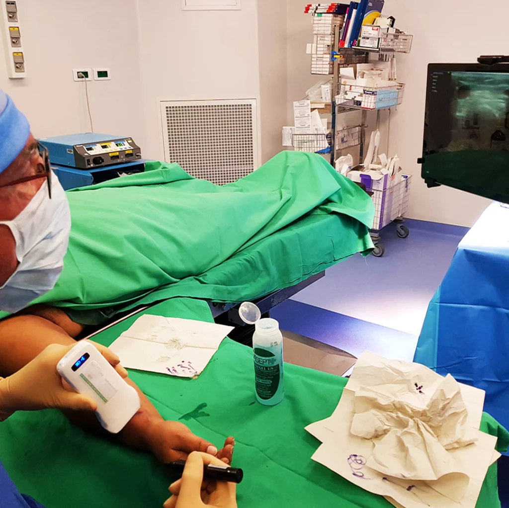
Linear Array Probe (4-15 MHz)
Ideal for facial aesthetics, superficial vasculature mapping, and flap planning. High-frequency resolution for delicate structures.
Curvilinear Probe (2-5 MHz)
Doppler with PW/CW Modes
Essential for mapping blood flow in flap surgeries and avoiding vascular complications.
Needle Guidance Overlay
User-Friendly Software
User-Friendly Software
What You Can Do with SonoMaxx Ultrasounds in Your Plastic Surgery Practice
Visualize filler depth, detect vascular structures, and ensure symmetry during treatments—all with handheld clarity and control.
SonoMaxx delivers detailed, real-time visualization of tissues and vessels—making procedures safer and outcomes more predictable.
- Guide dermal fillers with precise vascular mapping to reduce intravascular injection risks
- Assess fat graft distribution and retention for balanced, symmetric results
- Locate scar tissue and altered planes in revision surgeries to plan confidently
- Scan for seromas and hematomas during post-op visits without referring out
- Visualize perforators and vascular supply before flap-based reconstructions
- Build trust with patients by showing live imaging during consultations
Our handheld ultrasound devices are the trusted companion of plastic surgeons, aesthetic specialists, and reconstructive surgeons worldwide.


Why Leading Plastic Surgeons Choose SonoMaxx Handheld Ultrasounds
Engineered for aesthetic and reconstructive work, SonoMaxx provides high-definition imaging, portability, and intelligent tools tailored to your workflow.
01.
Exceptional Clarity for Facial Anatomy
Our high-resolution linear probes deliver shar pvisualization of dermal and subdermalstructures, ideal for skin and soft tissue
evaluatio
02.
Built-In Tools for Filler Safety
Scan in real time to map vessels, confirm filler location, and ensure even distribution withscan-labeling and comparison tools
01.
Trajectory-Guided Needle Insertion
Real-time overlay shows where your needle is headed—reduce pneumothorax and increase first-pass success on nerve and central line placements.
03.
Lightweight and Wireless
Slim, cordless design for hygienic, fast workflows-no tangle, no clutter, and perfectfor small treatment rooms.
04.
Designed for Aesthetic Workflows
Includes preloaded face sets, annotation tools,and side-by-side scan review for consultations and follow-up assessments.
05.
Boost Client Confidence and Retention
Show clients what’s beneath the surface, trackresults over time, and make consultations educational and visual.
06.
Trusted by Leaders in Aesthetic Medicine
Used globally by top cosmetic physicians and educators for evidence-based injection techniques and patient safety.
Trusted by 1000+ Plastic Surgeons, Aesthetic Experts & Reconstructive Teams
Free Software, Unlimited Scans
SonoMaxx’s handheld ultrasounds come with powerful software that gives you unlimited features at no extra cost. Enjoy free DICOM and pulsed wave doppler, plus a user-friendly interface on iOS, Android, and Windows—so you can focus on delivering top-quality care without any software fees.

Full Platform Compatible, Lifetime Free Upgrades



Capture Every Detail, Guide with Precision, and Share with Ease
Improve your ultrasound workflow with our all-in-one solution. Experience precision, simplicity, and efficiency in every scan.
17+ Specialized Presets
Convex / Phased Probe
Abdomen Gynecology Obstetric Cardiac
Urology Kidney Lung
Linear Probe
Thyroid Skin Small Parts Pediatrics
Vascular Carotid Breast MSK
Nerve Vessel Flow

B Mode

M Mode

Color Doppler

Power Doppler

Pulsed Wave Doppler
27 Automated Measurements for Precise Diagnostics
Simplify your work with a complete set of tools for fast, real-time assessments.
B-Mode
- Length, area/circumference, angle, trace, distance
- Gestational age (CRL, BPD, GS, FL, HC, AC)
- Estimated fetal weight (EFW) using BPD and FL
B+M Mode
- Heart rate, time, distance
- Left Ventricular Internal Diameter (LVIDd, LVIDs)
- Ejection Fraction (EF), Stroke Volume (SV)
B+PW Mode
- Velocity, heart rate, S/D ratio, depth
- Automatic vessel flow measurement
- Peak Systolic Velocity (PS), End-Diastolic Velocity (ED)
- Time-Averaged Maximum Velocity (TAMAX), Time-Averaged Mean Velocity (TAMEAN)
- Pulsatility Index (PI), Resistive Index (RI)
- Vessel Diameter (DIAM), Flow (FL)

Record up to 1000 frames (≈165 seconds) of continuous motion to capture every subtle detail.
Enjoy precise in-plane and out-of-plane guides to boost your confidence during interventions.
Save and share your scans effortlessly in JPEG, MP4, PNG, or DICOM formats, seamlessly integrating with your workflow.
How can we help you?
Do you need help finding a solution or choosing the right scanner for your practice? We’re here to help.
Will be used in accordance with our Privacy Policy
FAQ
How accurate is SonoMaxx Ultrasound for visualizing small-flow skin vessels?
All SonoMaxx devices come equipped with advanced Power Doppler technology. This makes it highly effective for assessing soft tissue infections, vascular malformations, or skin perfusion—especially in sensitive cases where clinical presentation alone is not enough.
Are SonoMaxx ultrasounds useful for breast implant assessments or other major augmentation surgeries?
Absolutely. SonoMaxx ultrasound systems with curvilinear probes can image structures ranging from superficial muscle layers to deeper tissues. This includes checking implant positioning, detecting capsular contracture, or identifying ruptures—all without needing an MRI.
Do SonoMaxx Ultrasound devices support video capture or just still images?
Both. You can capture high-resolution still images or record short, real-time video loops during your scan. This feature is particularly valuable when documenting movement—like joint motion or vascular flow—or when reviewing and sharing dynamic findings with colleagues, patients, or caregivers during follow-ups or telemedicine consults.
How do I store and share patient scans with SonoMaxx Ultrasounds?
Scans are saved securely to your iOS or Android device in EMR-compatible formats. Annotations and notes are preserved, and files can be shared instantly through PACS, DICOM, or cloud-based systems. You can also send scans via common communication apps, making documentation and consultation fast and fully integrated with your workflow.