Rib Fracture in POCUS: Seeing Beyond What Appears on X-ray
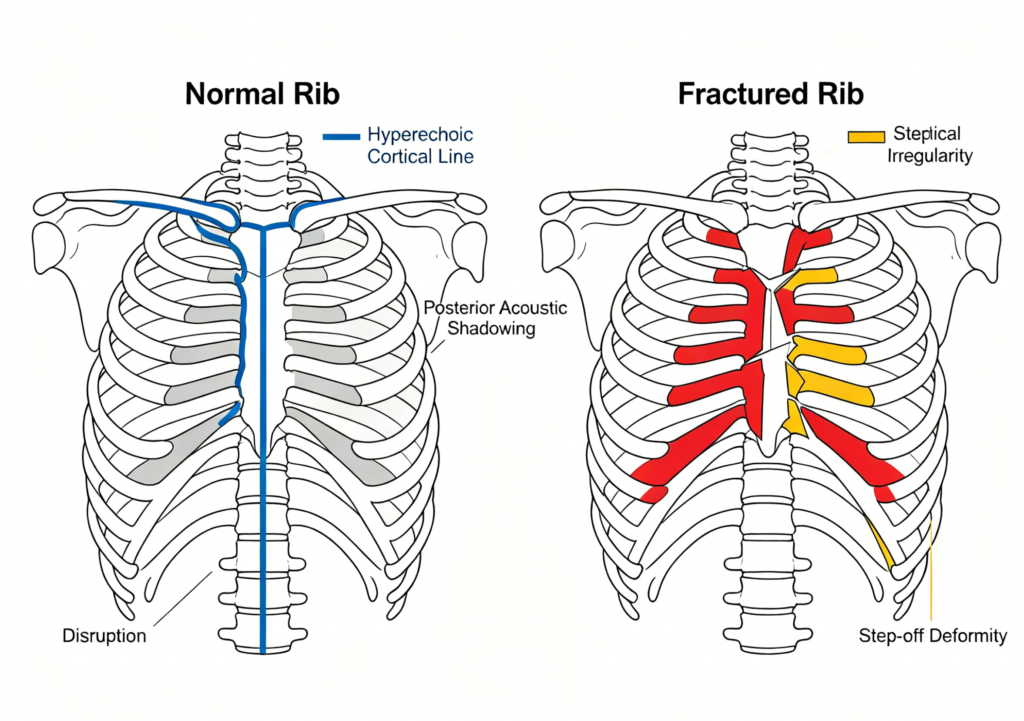
This image demonstrates a rib fracture identified using Point-of-Care Ultrasound (POCUS). On ultrasound, a normal rib appears as a continuous hyperechoic cortical line with posterior acoustic shadowing. A fracture may present as cortical disruption, irregularity, or a visible step-off deformity.
How do we assess a rib fracture using POCUS?
A high-frequency linear transducer is typically preferred for evaluating superficial structures. Scanning is performed directly over the area of maximal tenderness while following the rib along its course.
Key scanning points:
• Scan an adjacent non-tender area first to recognize normal anatomy.
• Follow the rib rather than focusing only on the painful point.
• Evaluate in at least two imaging planes.
• Use sonopalpation to correlate tenderness with ultrasound findings.
Beyond detecting the fracture itself, POCUS allows rapid assessment for associated findings such as:
• Pneumothorax
• Pleural effusion
• Local hematoma
• Adjacent soft tissue injury
POCUS provides immediate bedside information, but findings should always be interpreted together with clinical history, physical examination, and overall clinical judgment.